Our Services
At Retina Wellness & Surgery, we diagnose and treat a variety of common eye ailments with the utmost care, so that you can get back to living life to the fullest.
")
Some important things to know in preparation for your first visit:
-
Please plan on 1.5 to 2 hours for your first visit.
-
Bring sunglasses (rain or shine) because your eyes will be dilated. If possible, arrange for someone to drive you home.
-
If your insurance requires co-payments, we accept cash, check, credit, debit cards or digital payments such as Paypal, Venmo, Zelle and Cash App.
Other Items you'll need:
-
Your current medical insurance card(s).
-
Picture ID such as a driver's license.
-
A list of all medications you are currently taking.
-
If your insurance requires authorization for you to see a specialist, please bring an insurance referral which is separate from your insurance card. This is typically obtained from your insurance company or your primary care physician. If you have any questions or concerns about insurance or billing, please contact our office at 540-920-2020.
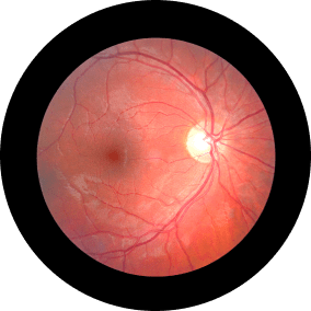
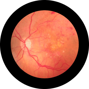
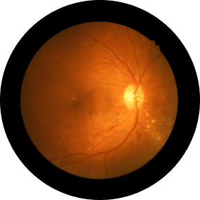
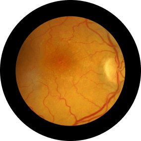
Diagnosis
Flashes/Floaters/
Shadows
Sudden onset of floaters, cobwebs, blurred vision, flashes of light or shadows especially in dim environment or with eye movement.
Macular
Degeneration
Gradual loss of the central
vision or no symptoms.
Diabetic
Retinopathy
Blurred vision, floaters,
distortion of vision.
Retinal Vascular
Occlusions
Painless acute vision loss of one eye, sudden blind spot in the visual field of one eye.
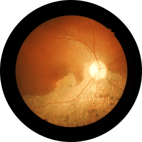
Epiretinal Membrane/
Macular Hole
Decreased vision, distorted
vision, or no symptoms.
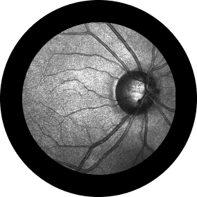
Uveitis
Pain, redness, light sensitivity,
decreased vision, vision
distortion, or floaters.
Treatment
")
Laser
With laser surgery (photocoagulation), your ophthalmologist uses a laser to make small burns around the retinal tear. The scarring that results will seal the retina to the underlying tissue, helping to prevent a retinal detachment. Retinal Laser Surgery is also employed in the treatment of retinal holes, lattice degeneration, central serous retinopathy, diabetic retinopathy, retinal vascular diseases, and other abnormalities.
Intravitreal Injections
An intravitreal (pronounced in tra VIT re al) injection is a procedure to place medication directly into the space in the back of the eye called the vitreous cavity, which is filled with a jelly-like fluid called the vitreous humor gel.
Intravitreal injections are performed in the office, often with the patient reclined in a chair. First, the eye and eyelids are anesthetized using drops or gel so the injection doesn’t hurt. Sometimes a small numbing injection may be given.
The eye and the eyelids are then cleaned, usually using povidone-iodine, a yellow solution which is very effective at killing bacteria that live around the eye. An eyelid speculum is often used to keep the eyelids open during the procedure. Once the eye is prepped for injection, you will be asked to look in a particular direction depending on the location of the injection while the medicine is injected through the pars plana (the white part of the eye) with a very small needle.
Typically, patients feel pressure, with little or no pain during the injection. After the injection, the speculum is removed and the eye is cleaned.
")
")
Surgery
Vitreoretinal surgery has advanced significantly over the past 15 years due to innovations in equipment, laser technology, fiber optics, and the introduction of vitreous substitutes such as purified silicone oil and perfluorocarbon gases. These tools have enabled the vitreoretinal surgeon to improve visual function in a variety of diseases.
The current retinal and vitreous treatments have resulted in improved management of postoperative infections following cataract and glaucoma surgery. Removal of retained intraocular foreign bodies has been improved as well as the management of complicated cataracts, dislocated intraocular lenses, and dislocated cataract material into the vitreous cavity. Diagnostic vitrectomy has improved the diagnosis of rare intraocular tumors. The surgical management of proliferative diabetic retinopathy, diabetic macular edema, and vascular occlusive disease has improved the visual function of many patients who were once deemed inoperable and destined for blindness.
The surgical management of macular abnormalities such as full-thickness macular holes, macular epiretinal membranes, and selected forms of macular degeneration is now possible. Many of these conditions may be treated with small-gauge or “suture-less” surgery.